Address: 155 State St Hackensack NJ 07601

WALK-IN X-RAY SERVICE AVAILABLE

NPI# 1285006791
Archives

Author's Posts

A closed cylindrical MRI scanner can provide nice MRI images in right patient or right clinical situation. Here at Affinity Imaging in Hackensack NJ we have a great 3.0T Machines. However there are many situations where Open MRI scanners can provide great images and help people who have a fear of confined spaces. In some cases an open MRI examination can even provide greater patient safety because of access and speed of the scan.

At Affinity Radiology in Hackensack, NJ we have an Open MRI that is able to provide imaging evaluation of patients who are physically too large to be comfortably placed within a conventional cylindrical MRI scanner.

The Open sides of an open MRI allow patients to see the room surrounding the scanner to minimize claustrophobia. There is also easy access to a friend or family member to comfort the patient or to provide necessary physical or medical support during an MRI examination.


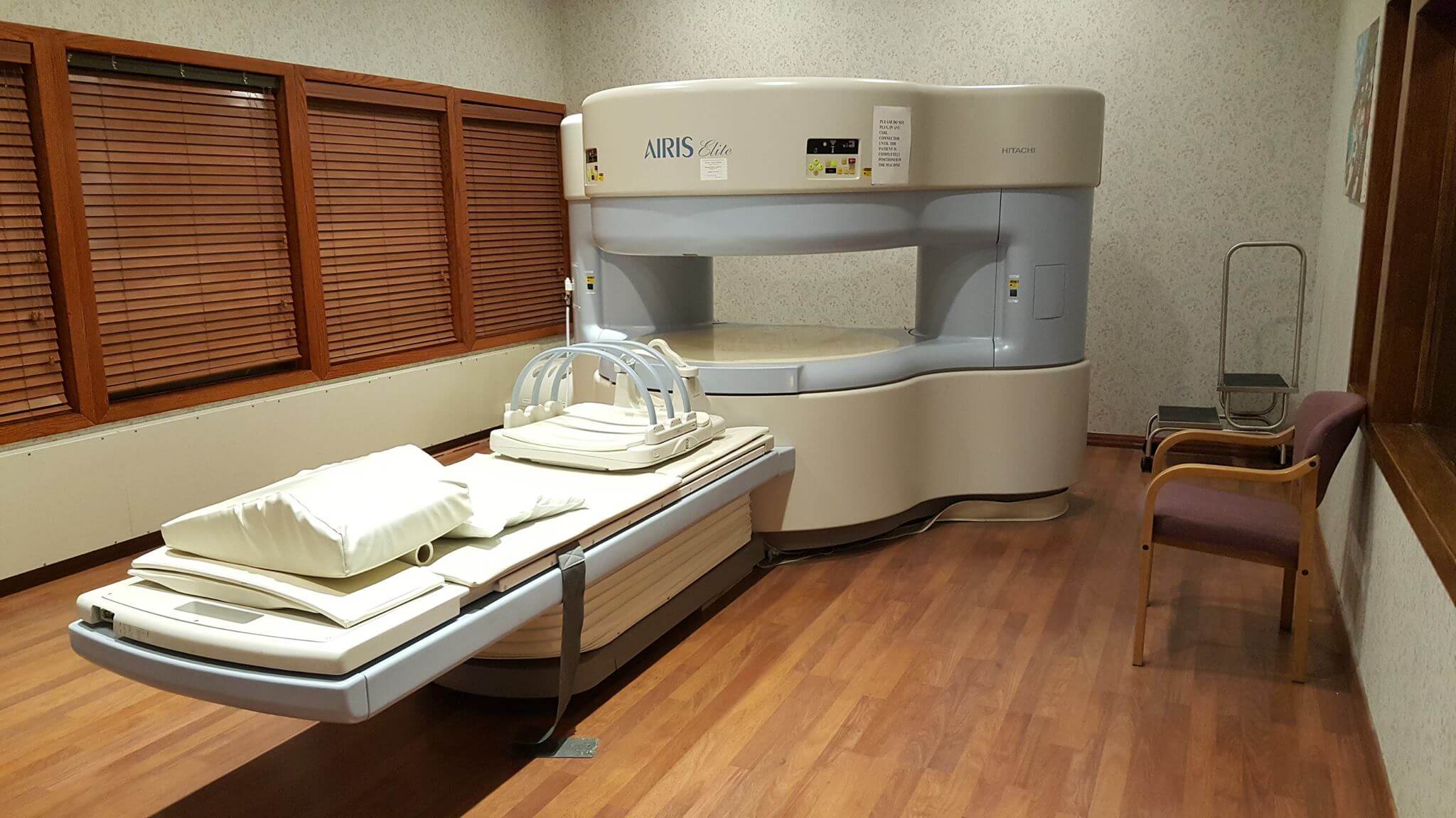
The wide open design of an Open MRI allows the critical body parts of a patient to be positioned near the center of the magnet where the best images of the body part are obtained. A closed cylindrical shaped MRI does not allow all body parts to have the best position in the magnet and larger patients may have an even greater problem with optimal positioning of critical body parts within the MRI scanner.

Open MRI scanners produce less noise than closed MRI scanners. Loud noises of closed MRI examinations can be uncomfortable and in some cases can damage hearing, so closed MRI scanners may require ear plugs to protect hearing and minimize discomfort. An open MRI examination may not require ear plugs.

Many patients have metallic objects within their bodies from previous surgery. Artifacts from metal implants can greatly reduce the quality of MRI images. However the lower magnetic field strength open MRI scanners produce less artifact from metallic implants than higher magnetic field closed MRI scanners, so patients with metallic implants may obtain greater benefit from an open MRI examination.

The rapidly changing magnetic fields and radio waves used by MRI generate heat within metallic implants within the body or metal attached to the body, including some dyes within tattoos or mascara. This heating effect is decreased and sometimes eliminated by the lower magnetic field strength open MRI scanners.

Lower field strength open MRI scanners have greater contrast between different types of body tissues, so it may be easier to find abnormal body tissues with an open MRI even if there is less spacial detail within the body tissues. This greater natural tissue contrast provided by an open MRI decreases the need for the injection of chemical contrast agents into the veins of the patient.

If you or a loved one has any questions about our MRI either Open or Closed call us at 201-968-5544 and a friendly staff member can help you with your questions. We participate with all major medical insurance companies and Medicare and can schedule you for a same-day appointment if needed.

Hackensack Radiology Center offers Open MRI Scans

Patients who are larger, or suffer with even mild claustrophobia, may find it difficult to tolerate long scan times inside the machine. Hackensack Radiology Center uses OPEN MRI Technology to accommodate these patients and help overcome their discomfort. The OPEN MRI is a machine that is open on the sides rather than a tube closed at one end, so it does not fully surround the patient. Newer OPEN MRI Technology, like the ones used at Hackensack Radiology Center, provides high quality images.
Continue reading Open MRI Scan

About Magnetic Resonance Imaging

MRI is a non-invasive imaging technology that produces three dimensional detailed anatomical images without the use of damaging radiation. It is often used for disease detection, diagnosis, and treatment monitoring. It is based on sophisticated technology that excites and detects the change in the direction of the rotational axis of protons found in the water that makes up living tissues.
Continue reading Magnetic Resonance Imaging

About DEXA

Dual-energy X-ray absorptiometry (DXA, previously DEXA) is a means of measuring bone mineral density (BMD). Two X-ray beams, with different energy levels, are aimed at the patient’s bones. When soft tissue absorption is subtracted out, the BMD can be determined from the absorption of each beam by bone. Dual-energy X-ray absorptiometry is the most widely used and most thoroughly studied bone density measurement technology.
Continue reading DEXA Bone Scan

About Ultrasound

Medical ultrasound (also known as diagnostic sonography or ultrasonography) is a diagnostic imaging technique based on the application of ultrasound. It is used to see internal body structures such as tendons, muscles, joints, vessels and internal organs. Its aim is often to find a source of a disease or to exclude any pathology. The practice of examining pregnant women using ultrasound is called obstetric ultrasound, and is widely used.
Continue reading Ultrasound

About PET/CT Scan

Positron emission tomography–computed tomography (better known as PET-CT or PET/CT) is a medical imaging technique using a device which combines in a single gantry system both a positron emission tomography (PET) scanner and an x-ray computed tomography (CT) scanner, so that images acquired from both devices can be taken sequentially, in the same session, and combined into a single superposed (co-registered) image. Thus, functional imaging obtained by PET, which depicts the spatial distribution of metabolic or biochemical activity in the body can be more precisely aligned or correlated with anatomic imaging obtained by CT scanning. Two- and three-dimensional image reconstruction may be rendered as a function of a common software and control system.
Continue reading PET/CT Scans

About Digital Mammography

Digital mammography is a specialized form of mammography that uses digital receptors and computers instead of x-ray film to help examine breast tissue for breast cancer.The electrical signals can be read on computer screens, permitting more manipulation of images to theoretically allow radiologists to more clearly view the results. Digital mammography may be “spot view”, for breast biopsy, or “full field” (FFDM) for screening.
Continue reading Digital Mammography

Book your appointment

Walk-In X-Ray Service Now Available

Call Us 201 968 5544
cancel payment

Online Bill Pay

A 3% CONVENIENCE FEE WILL BE ADDED

Need Help? PLEASE CALL OUR OFFICE AT (201) 968 5544

PayPal Acceptance Mark

cancel payment

Online Bill Pay

a 3% convenience fee will be added

Need Help? PLEASE CALL OUR OFFICE AT (201) 968 5544

PayPal Acceptance Mark

cancel payment

Online Bill Pay

all fields required

Need Help? PLEASE CALL OUR OFFICE AT (201) 968 5544